Monday, April 22, 2024

Case of the Week 746

Welcome back to our microfilariae block from Idzi Potters and the Institute of Tropical Medicine, Antwerp! We are down to the last TWO specimens. This week's case is from a skin specimen - both skin samples AND a subcutaneous nodule (removed from the upper arm). The patient is a young man from Brazil.

Here is the nodule:

And here are the skin scarification specimens (read more about scarification vs. skin snips HERE):

Carazzi stain:


Giemsa stain:

The microfilariae are approximately 300 micrometers in length. What is your diagnosis?

Monday, April 15, 2024

Case of the Week 745

We have moved onto microfilariae found in SKIN specimens - what fun! There are only a couple to choose from. Can you tell which one this is? The microfilariae are approximately 200 micrometers long.

Carazzi stain


Giemsa stain:


Sunday, April 14, 2024

Answer to Case 745

 Answer to the Parasite Case of the Week 745: Mansonella streptocerca microfilariae

Florida Fan and Anonymous pointed out the small size and the source of skin as key features for  diagnosing Mansonella streptocerca infection. Idzi also noted on LinkedIn that "A nice little detail is the etiology of the name “streptocerca”! It is derived from “strepto” meaning “twisted chain” and “cerca” meaning “tail”. The row of single round nuclei in its tail do resemble a chain (or necklace) in my opinion. The prefix “strepto” is also used for streptococcus - which indeed also forms “chains”…" What a fun insight!

Here is an image from this case that nicely highlights these features:

Thank you all for sticking with us for this fun series of microfilariae. We have TWO left for you, so stay tuned!
 

Monday, April 8, 2024

Case of the Week 744

 We are in the home stretch for the microfilariae! Can you believe that we have just a few more to go? (Unless I decide to repeat some ๐Ÿ˜‰)  This week's case shows microfilariae that are approximately 200 micrometers long. The patient is a resident of Brazil and has moderate peripheral eosinophilia. He is otherwise asymptomatic. 

Carazzi stain (Knott's concentration):


Giemsa stain (thick blood film):


Sunday, April 7, 2024

Answer to Case 744

 Answer to the Parasite Case of the Week 744: Mansonella perstans microfilariae.

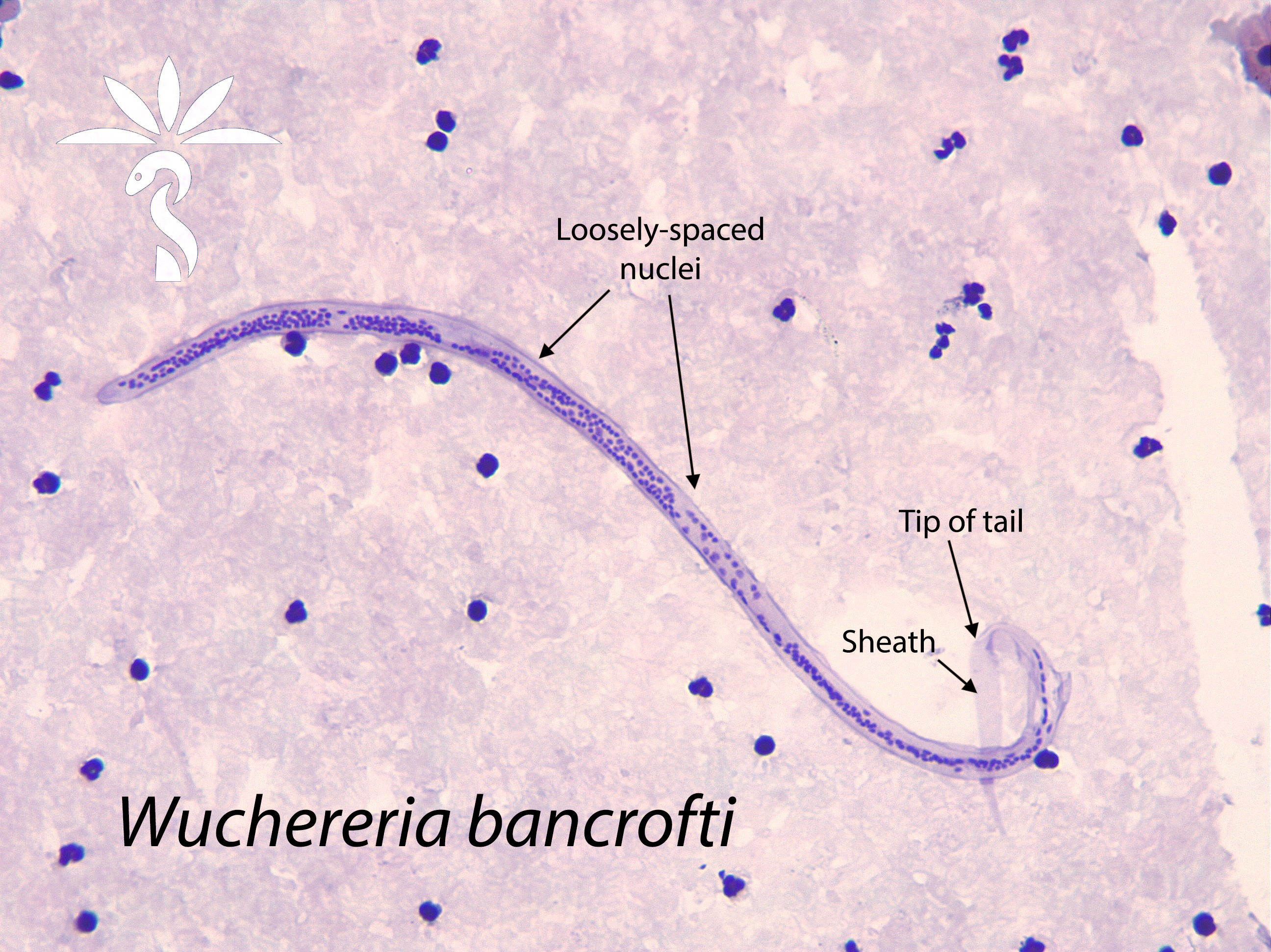
As described by Florida Fan, "This is a rather small microfilaria, its width is only about half the diameter of the surrounding neutrophils. The Carrazi stain [a hematoxylin-based stain] did not show a sheath either. As such, we can definitely rule out all the sheathed and large microfilaria. We know that we are dealing with Mansonella species. The tail of this Mansonella is not curved , this allows us to eliminate Mansonella streptocerca (strepto = curved, cerca = tail) [and also the source is not tissue]. The tail is also not pointed, this rules out Mansonella ozzardi. We only have one left with a blunt tail: Mansonella perstans which persists." 

This image nicely shows all of these features:

You have all done a great job learning to differentiate the small, unsheathed blood microfilariae (i.e. Mansonella perstans and M. ozzardi) from the larger, sheathed microfilariae. 

Of course, co-infections can occur, and the following is a stunning photograph of M. perstans and Loa loa co-infection. I think we can all appreciated that the Loa loa microfilaria is the top based on its larger size and sheath. 

Thank you for these outstanding cases, Idzi!

Next week we will finish up with the tissue microfilariae. Will you be able to tell them apart?


Monday, April 1, 2024

Parasite Case of the Week 743

Welcome back for more microfilariae from Idzi Potters and the Institute of Tropical Medicine, Antwerp. The following microfilariae were seen in Giemsa-stained thick blood films from a man living on Alor Island, Indonesia. They measure approximately 305-315 micrometers in length. 


What is your identification? What is your primary differential?


Sunday, March 31, 2024

Answer to Case 743

 Answer to the Parasite Case of the Week 743: Brugia timori microfilariae. This was one of the tougher cases, but everyone did a great job narrowing the differential to Brugia. As noted by Florida Fan and Anonymous, we can immediately rule out Mansonella species based on the small size (length and diameter) of the microfilariae. Florida Fan also notes that the sheath is visible, confirming that we are dealing with Loa loa, Wuchereria bancrofti or the Brugia genus. 

He then details his method for coming to an exact diagnosis: "First the column of nuclei is so compact that we can rule out Wuchereria bancrofti, also the nuclei go to the end of the tail. Yet, the two terminal nuclei are distinctly separated from the nuclei column, this rules out any chance of being Loa loa. Now we have the Brugia genus left, but where is the pink sheath pertinent to Brugia malayi when stained with Giemsa stain? As such, we eliminate Brugia malayi. By the elimination process, we’re left with its cousin Brugia timori, the sheath of this one does not stain pink with Giemsa stain. Now let’s check the map of the lesser Sundae island, the Alor island is right North of Timor."

I will also point out that the head space is longer than that of B. malayi and there are a larger number of single-file nuclei going towards the tail. These features are more subtle and can be difficult to appreciate. I've done my best to point out some of them in the image below.

Thank you all for sticking with us on our microfilariae journey. The microfilariae can be very challenging to identify, and are rare in many parts of the world, so our lab staff may not have the opportunity to examine these organisms outside of their EQA/PT schema. 


Monday, March 25, 2024

Case of the Week 742

This week's case features another beautiful example of microfilariae in blood. The patient is from sub-Saharan Africa and presents with chronic swelling of his left leg. The microfilariae measure approximately 270 ยตm in length. 

Carazzi stain (Knott's concentration):


Giemsa Stain (thick blood film)

What is your identification? What time should blood be collected for this examination?

Sunday, March 24, 2024

Answer to Case 742

 Answer to the Parasite Case of the Week 742: Wuchereria bancrofti microfilariae.

I really enjoyed reading the comments on this case. FloridaFan provided this excellent description of his approach to microfilariae identification: "First the width of the worm is about the same as that of the surrounding neutrophils. Second, its length is greater than 200 micrometers. Third, it has a sheath. Now we know we are dealing with Loa loa, Wuchereria, or the Brugia ones, not with any little pesky Brugia. The next consideration is that the column of nuclei is continuous, the terminal nuclei are not separate from the immediate anterior nuclei. This eliminates the possibility of the Brugia malayi and timori, over that these are “far fetched” geographically. We are left with two candidates Loa loa and Wuchereria bancrofti. Though the tail is not so obvious, the Carrazi stain did show that the nuclei column terminates well short from the end of the tail. This rules out Loa loa. The only culprit left is Wuchereria bancrofti.

An anonymous commenter also noted that "Its nuclear column is relatively loose, and individual nuclei can be visualized throughout the column. The sheath is does not stain pink with Giemsa stain (as in Brugia malayi)."

These features can be nicely visualized in the photo of the Carazzi-stained Knott's concentration. Note that the Carazzi stain is a hematoxylin-based stain (rather than conventional Giemsa) that helps to demonstrate the sheath. It is a very useful stain to have!

Thanks again to Idzi Potters and the Institute of Tropical Medicine, Anwerp, for this beautiful case.


Monday, March 18, 2024

Case of the Week 741

Wow, we are already on our 3rd filarial case! The following lovely case from Idzi Potters and the Institute of Tropical Medicine, Antwerp, is another microfilaria found in blood. 

The patient is a middle aged male farmer from Central America who was noted to have mild eosinophilia on routine complete blood count. He is otherwise asymptomatic. The microfilariae measure approximately 175 micrometers in length. What is your identification?